
The terms Angiography vs Angioplasty often come up in conversations about heart and vascular health. This can lead to confusion because they sound similar and involve comparable procedures. However, there are key differences in their purpose, execution, and outcomes. One procedure is for diagnosis(Angiography), while the other is for treatment (Angioplasty). Understanding this distinction is important for patients and their families as they navigate cardiovascular care, especially when dealing with conditions like coronary artery disease (CAD) or peripheral artery disease (PAD).
This article aims to clarify the roles of each procedure, including what they are, why they are done, and how they work. We will discuss the difference between viewing blockages (angiography) and physically clearing those blockages (Angioplasty). This knowledge will help readers have informed discussions with their healthcare providers about their diagnosis and treatment plans for better blood flow and improved heart health.
The Fundamental Purpose: Diagnosis Versus Treatment
Angiography—The Diagnostic Tool
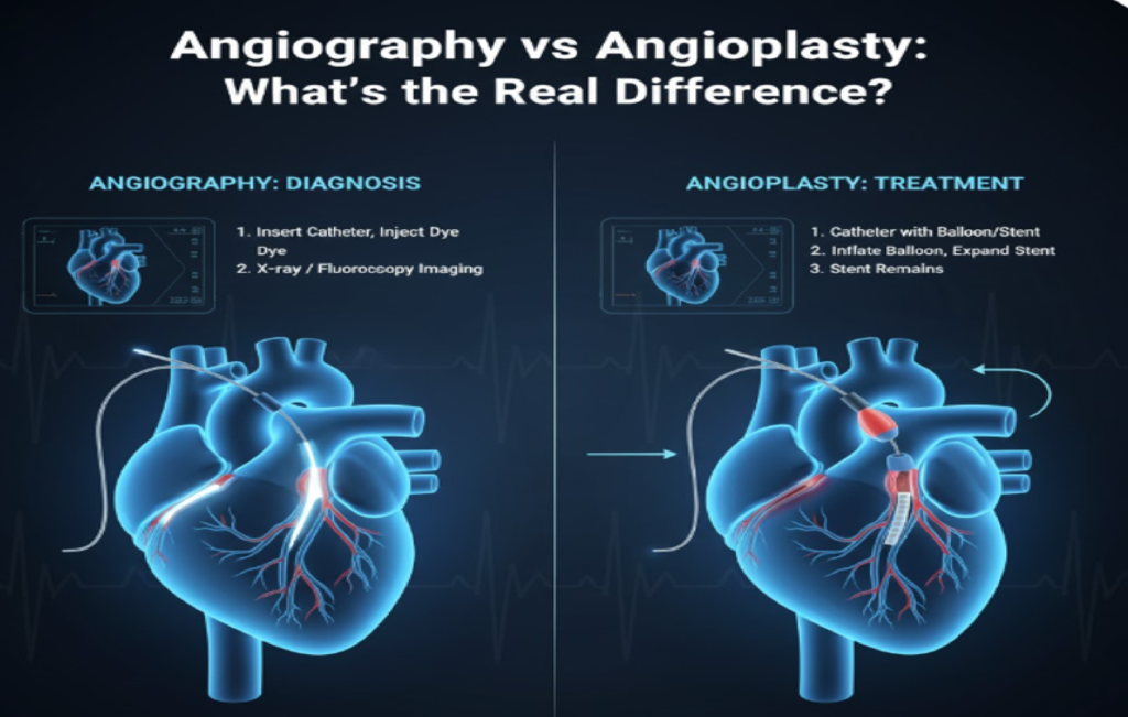
The main difference between angiography and angioplasty is their clinical goals. Angiography is a diagnostic procedure. Its purpose is to visualize blood vessels and find any blockages, narrowing (stenosis), or aneurysms. It works like a detailed X-ray map of the arteries, showing the location and severity of any vascular disease. Angiography provides information, not a cure, and serves as the first step to confirm a diagnosis suggested by non-invasive tests like a stress test or echocardiogram.
Angioplasty – The Therapeutic Procedure
In contrast, Angioplasty is a therapeutic procedure. Its goal is not to look, but to fix. This intervention physically reopens narrowed or blocked arteries, restoring blood flow to downstream tissues, typically the heart muscle. Angioplasty often follows an Angiography when a significant blockage is found, turning a diagnostic session into a life-saving intervention called percutaneous coronary intervention (PCI).
Angiography Explained: The Diagnostic Roadmap
How Angiography Works
Angiography, also known as arteriography, is a precise imaging technique that creates detailed pictures or angiograms of blood vessels. The procedure usually takes place in a catheterization laboratory, or “cath lab,” where a thin, flexible tube called a catheter is inserted into an artery, often in the wrist or groin. The catheter is then guided through the vascular system to the area of interest, such as the coronary arteries around the heart. Once positioned, a special iodine-based contrast dye is injected through the catheter.
What Patients Can Expect
This dye blocks X-rays, making the blood vessels visible under fluoroscopy, a live X-ray imaging method. As the dye flows through the arteries, the physician can observe and record the flow in real time. If a significant blockage exists, the flow of the dye will slow down or stop, outlining the diseased segment of the artery and providing the physician with a critical roadmap for future procedures.
Angioplasty Explained: The Therapeutic Intervention
Balloon Expansion Process
Angioplasty, typically performed during Percutaneous Coronary Intervention (PCI) when addressing coronary arteries, aims to clear arterial blockages. The procedure uses the same entry points and catheterization techniques from the . Once the has identified a flow-limiting blockage, the physician threads a special guidewire across the narrowed section of the artery. A small balloon-tipped catheter is then advanced over this guidewire and positioned within the blockage. The balloon is inflated to high pressure, compressing the plaque against the artery walls, expanding the artery’s diameter and restoring blood flow.
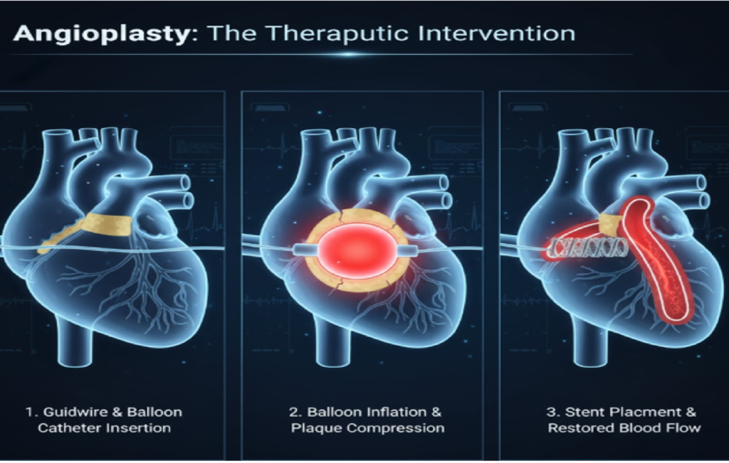
This expansion of the vessel gives the procedure its name: Angio (vessel) + plasty (shaping). After the inflation, the balloon is deflated and removed. Angioplasty provides immediate relief of symptoms like chest pain and helps prevent heart muscle damage during a heart attack.
The Role of Stenting: Essential Component of Modern Angioplasty
While angioplasty primarily refers to the use of a balloon to open a blocked vessel, modern practices usually involve placing a stent afterward. A stent is a small, metallic mesh tube that acts as a support to keep the widened artery open. After the balloon dilation pushes the plaque aside, the stent, mounted on a second balloon catheter, is delivered to the site. The balloon is inflated again to expand the stent against the vessel wall.
The balloon catheter is then pulled out, leaving the stent in place to maintain the artery’s wider diameter and lower the risk of it closing off again (restenosis). Most stents used today are Drug-Eluting Stents (DES), which slowly release medication to prevent excessive tissue growth that can cause restenosis. Thus, while balloon angioplasty and stent placement are technically different, the combination is regarded as the standard for most current revascularization procedures.
Comparing Patient Experience and Recovery
Duration and Comfort
The experience for a patient undergoing Angiography is straightforward: it involves catheter insertion, contrast injection, and a series of X-rays. This typically lasts between 30 minutes to an hour. The patient is usually awake but sedated, and they often feel a warm flush when the contrast dye is injected. The risks are minimal and primarily relate to the catheter insertion site and potential allergic reactions to the dye. Angioplasty is more involved and time-consuming, often lasting between 1 to 3 hours, especially if multiple stents are placed.
Risks and Complications
It requires precise handling of balloons and stents across the blockage. Though both procedures take place in the cath lab and start similarly, the therapeutic nature of means it usually takes longer, uses more contrast dye, and has a higher potential for complications. Recovery times differ, too: angiography patients often go home the same day, while those who undergo
Comparing Risks and Complications: A Matter of Intervention
While both Angiography and Angioplasty are minimally invasive, catheter-based procedures, their risk profiles differ significantly because one is solely diagnostic and the other is therapeutic. Angiography carries lower risks, mainly centered around the use of the contrast dye and the catheter insertion site. Minor complications often include bruising, soreness, or bleeding at the wrist or groin access point, and a small risk of a mild allergic reaction to the dye. A rare but important risk is contrast-induced nephropathy (kidney damage), particularly in patients with pre-existing kidney issues. Angioplasty, being an intervention, inherently carries a higher risk of major complications. These include an increased risk of bleeding, particularly from the artery being manipulated, and
potential damage to the coronary artery itself, which in very rare cases can necessitate emergency bypass surgery. There is also a small risk of acute closure of the stented artery due to blood clots forming, which is why aggressive antiplatelet therapy is essential post-procedure. Overall, while both procedures are generally safe, the complexity of opening a blocked artery means Angioplasty requires stricter post-procedure monitoring and carries a statistically greater chance of a serious event, though the mortality rate remains very low, especially in experienced centres.
Beyond the Heart: Peripheral and Renal Applications
Though Angiography and Angioplasty are most commonly used for coronary arteries, these techniques are also applied throughout the vascular system. For example, in cases of Peripheral Artery Disease (PAD), maps blockages in the arteries of the legs. If significant narrowing is found that affects blood flow and causes symptoms like leg pain or non-healing wounds, and stenting can restore circulation to the limbs. Similarly, Renal Artery Stenosis, which narrows the arteries that supply the kidneys, can lead to high blood pressure and kidney failure.
Renal Angiography is used for diagnosis, followed by renal Angioplasty and stenting to open the blocked artery, aiming to lower blood pressure and protect kidney function. These applications illustrate that the core idea—
Conclusion: Choosing the Right Procedure
Understanding the difference between Angiography and Angioplasty is fundamental to modern cardiovascular care: one is the diagnostic tool that identifies the issue, while the other is the treatment that resolves it. Angiography maps the presence, location, and severity of arterial blockages through contrast dye and X-rays. Angioplasty, typically with stenting, is the active treatment that uses a balloon and a permanent mesh scaffold to restore blood flow through narrowed arteries. Recognizing this distinction helps patients follow the step-by-step process of their care, from diagnosis to treatment.
Whether managing urgent heart attacks or chronic peripheral artery disease, these catheter-based procedures offer effective, less invasive options compared to open surgery. Knowing the roles of these procedures is the first step in making informed decisions about your vascular health. Do you have any specific questions about preparing for or recovering from either procedure?
Book a Heart Check-Up Today →
Early diagnosis through angiography and timely treatment with can significantly improve heart health and long-term outcomes.